Our Equipment
The Graham Lab uses an array of equipment to image the healthy and diseased brain. Within Sunnybrook Research Institute, we use the following:
MRI System
We use a 3.0 Tesla research-dedicated MRI system (Magnetom Prisma, Siemens) which was purchased through funds awarded to Sunnybrook Health Sciences Centre from the Canadian Foundation for innovation (CFI). This MRI system is highly versatile and the lab takes advantage of diverse imaging methods in various research projects. However, the principal imaging method that we use on the system is functional MRI (fMRI).
Briefly, the fMRI signal is created by changes in blood oxygenation, flow and volume that occur in small-scale vasculature in response to electrical activity in nearby neurons as they undergo mental processing. The technique has become a major tool in basic neuroscience to understand how brain activity is regionally and functionally distributed and is emerging as a tool that may have medical application in patients with neurological or psychiatric diseases.
Eye-Tracker
The EyeLink 1000 Plus is used by our team to analyze saccadic movement of the eyes, pupil dilation, and eye position - during behavioural testing that is conducted outside as well as inside the MRI system.
Image taken from SR Research (https://www.sr-research.com/eyelink-1000-plus/)
Electroencephalography
Electroencephalography (EEG) is the study of the electrical activity within the brain. Using EEG in our studies adds another layer to our understanding of brain activity. The lab has two EEG systems available:
- A high fidelity 64-channel system, purchased from Brain Products, and;
- A rapidly deployable 24-channel system for clinically focused EEG research, purchased from Advanced Brain Monitoring.
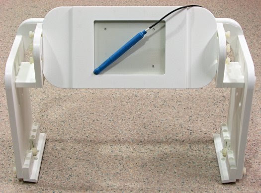
fMRI-Compatible Tablet
Our research into drawing and writing using fMRI has led to the development of a succession of fMRI-compatible tablets. For our applications, we prefer to use a commercially-available rear projection display viewed through a mirror on the head coil, although video goggles are also usable. Details of how the tablet works were published in our TECHNICAL PAPER and PATENT.
The tablet is touch sensitive and can be operated with fingers or any blunt object. Our favourite blunt object is a dual-function, force-sensitive stylus included with recent tablets (rev. 1.7+). The tablet plugs into a computer via USB and presents industry-standard interfaces that are compatible with a variety of software for programming fMRI tasks. The latest version includes numerous refinements to improve its ergonomics and robustness, as well as to meet stringent regulatory requirements for eventual medical device approvals.
We are striving to commercialize the tablet, but in the meantime we have already sold several tablets to labs around the world, on a cost-recovery basis. We are keen to continue development of the technology and to see more researchers use it. Please contact us () if you are interested in buying one for your lab.